At the Bhagwan Mahaveer Cancer Hospital & Research Centre (BMCHRC), our Radiology & Interventional Oncology represents the forefront of compassionate cancer care. We understand that a cancer diagnosis can be overwhelming, both physically and emotionally, which is why our dedicated team is committed to providing personalized and comprehensive care to every individual who walks through our doors.
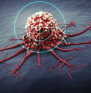
At BMCHRC, our Radiology and Interventional Oncology Services are integral components of our comprehensive cancer care approach. Led by a team of experienced radiologists and interventional oncologists, we offer advanced imaging technologies and minimally invasive procedures to diagnose and treat various cancers. Our state-of-the-art radiology department is equipped with cutting-edge imaging modalities, including MRI, CT scans, PET-CT scans, and ultrasound, enabling accurate and timely detection of tumors and metastases. Additionally, our Interventional Oncology Services provide minimally invasive treatments such as radiofrequency ablation, cryoablation, chemoembolization, and radioembolization, delivering targeted therapy directly to cancerous lesions while preserving healthy tissue. With a focus on precision, efficacy, and patient comfort, our Radiology and Interventional Oncology Services play a crucial role in optimizing cancer management and improving outcomes for our patients.

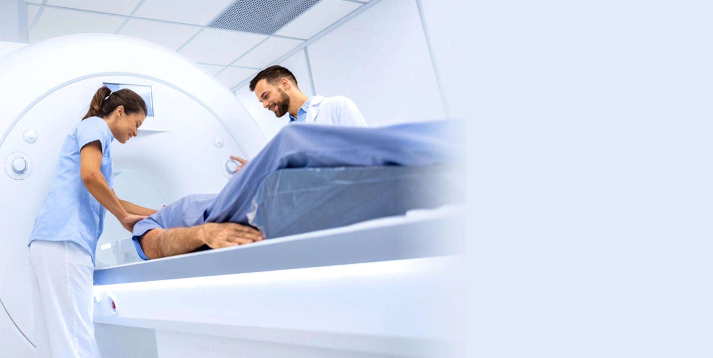
Magnetic Resonance Imaging (MRI) is a non-invasive medical imaging technique used to produce detailed images of the organs and tissues within the body. It is particularly useful for imaging soft tissues, such as the brain, muscles, and connective tissues. MRI is a crucial tool in modern medicine, providing critical information that can aid in diagnosis and treatment planning. In summary, MRI is a powerful diagnostic tool widely used in modern medicine due to its ability to produce detailed images of the body's internal structures without the risks associated with ionizing radiation.

Digital Radiography (DR) is an advanced imaging technology used in medical diagnostics that replaces traditional X-ray film with digital detectors. This system captures X-ray images directly onto a digital medium, which can then be processed, stored, and viewed on a computer. Digital radiography offers several advantages over conventional film-based radiography, including faster image acquisition, improved image quality, and enhanced workflow efficiency. Overall, digital radiography has transformed medical imaging by offering faster, more efficient, and higher-quality imaging capabilities, leading to improved patient care, diagnosis, and treatment outcomes.

Ultrasonography (USG) with Color Doppler is a medical imaging technique that combines traditional ultrasound imaging with Doppler ultrasound to visualize blood flow within the body's vessels and organs. This technology adds color to the ultrasound images, allowing healthcare professionals to assess blood flow patterns, direction, and velocity in real-time. USG with Color Doppler is widely used across various medical specialties for diagnostic purposes and to monitor conditions affecting blood circulation. USG with Color Doppler is a valuable imaging tool that aids in the diagnosis, monitoring, and management of vascular and circulatory disorders, cardiac conditions, obstetric and gynecological concerns, and various other medical conditions. Its real-time visualization of blood flow adds an important dimension to ultrasound imaging, enhancing the diagnostic capabilities across multiple medical specialties.
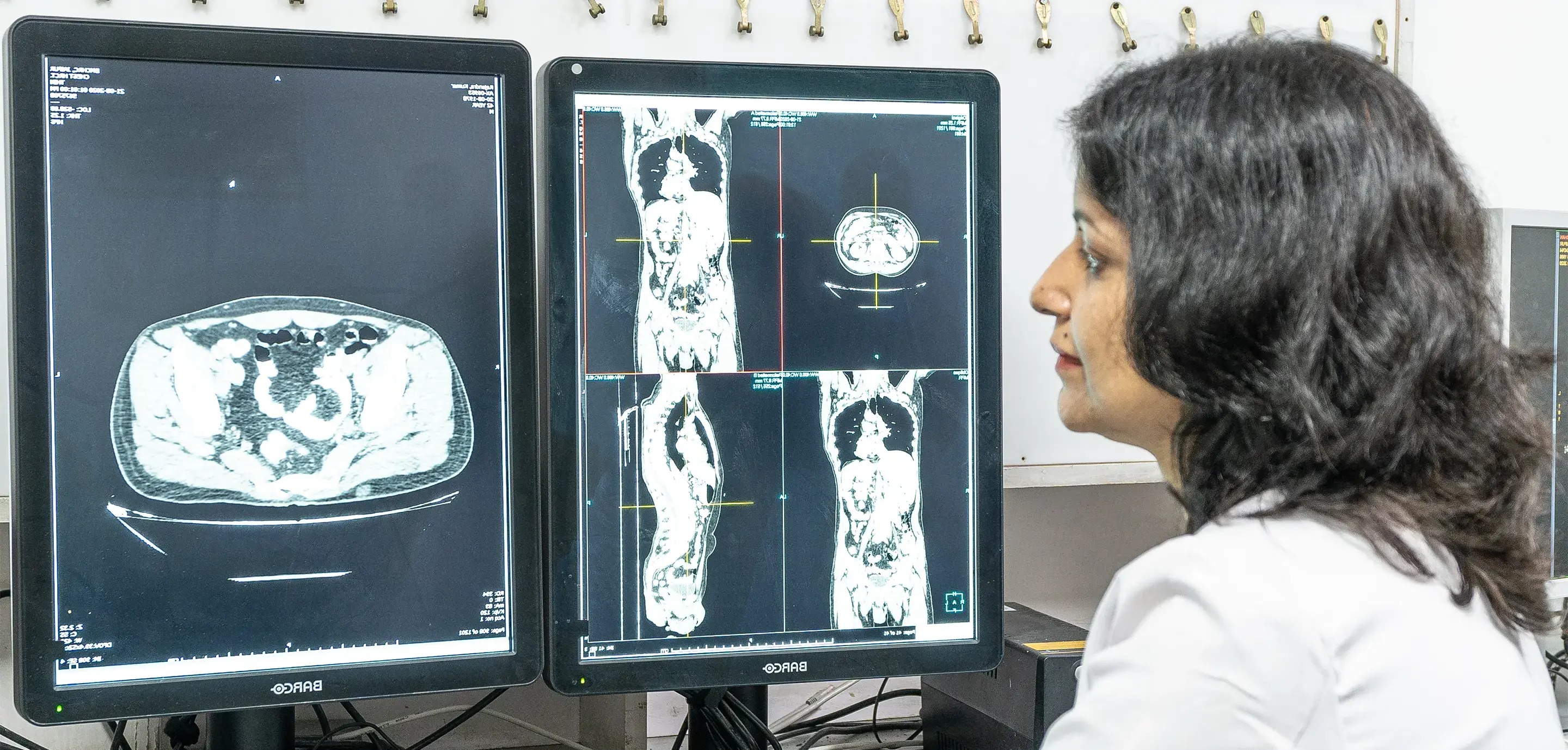
A Computed Tomography (CT) scan, also known as a CAT scan (Computerized Axial Tomography), is a medical imaging technique that uses X-rays and computer processing to create detailed cross-sectional images of the body. It provides a more comprehensive view than traditional X-rays and is particularly useful for diagnosing and evaluating a wide range of medical conditions, including injuries, diseases, and abnormalities. Overall, CT scans play a crucial role in modern medicine, providing detailed and comprehensive imaging for diagnosing and managing a wide range of medical conditions. They are particularly valuable in emergency settings, cancer diagnosis and staging, trauma assessment, and evaluating complex anatomical structures. However, healthcare providers must weigh the benefits of CT scans against potential risks and consider individual patient factors when ordering imaging studies.

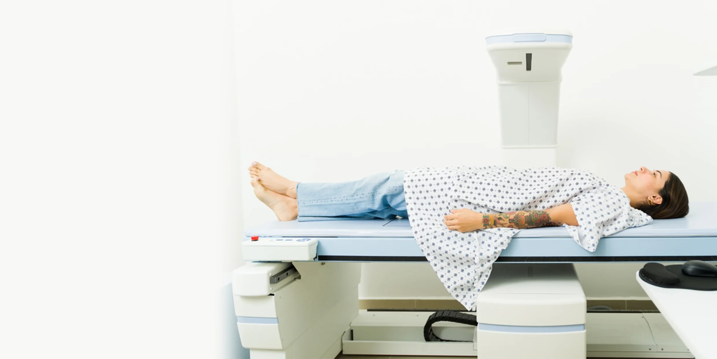
A Dual-Energy X-ray Absorptiometry (DEXA) scan is a medical imaging technique primarily used to measure bone mineral density (BMD) and assess bone health. It is a non-invasive and low-radiation procedure that helps in diagnosing osteoporosis, evaluating fracture risk, and monitoring changes in bone density over time. DEXA scans are commonly performed on the spine, hips, and sometimes the forearm. Overall, DEXA scans are valuable tools in assessing bone health, diagnosing osteoporosis, and guiding management strategies to reduce fracture risk and promote skeletal well-being.

Mammography with stereotactic biopsy is a diagnostic procedure used in breast imaging to evaluate suspicious breast lesions detected on a mammogram. It combines mammographic imaging with a minimally invasive biopsy technique called stereotactic biopsy, allowing for precise sampling of abnormal breast tissue for further evaluation. This procedure is commonly used to diagnose breast cancer and other breast abnormalities. Mammography with stereotactic biopsy is a valuable tool in breast imaging and the diagnosis of breast abnormalities, particularly for lesions that are not easily accessible for palpation or visual examination.
An "Orthopentogram" doesn't appear to be a standard medical term or imaging procedure. It's possible that you meant "orthopantomogram" (OPG), also known as a panoramic dental X-ray, which is a specific type of dental radiograph used to capture a wide-view image of the entire mouth, including the teeth, jaws, and surrounding structures. Overall, orthopantomograms (OPGs) play a significant role in dental diagnostics, treatment planning, and oral health assessments.
.webp)
Mobile X-Ray units are portable imaging devices used to perform X-ray examinations at the patient's bedside or in locations where traditional fixed X-ray machines are impractical or unavailable. These units are designed for flexibility, allowing healthcare providers to conduct diagnostic imaging studies on-site, such as in hospital wards, emergency rooms, intensive care units (ICUs), nursing homes, or outpatient clinics. Mobile X-ray units offer several advantages, including convenience, efficiency, and reduced patient movement for critical or immobile individuals. Despite these challenges, mobile X-ray units play a vital role in modern healthcare settings by providing accessible and timely diagnostic imaging services for patients who require immediate attention or cannot easily be transported to radiology departments.

Interventional Radiology (IR) is a medical specialty that uses minimally invasive procedures guided by imaging techniques such as X-rays, ultrasound, CT scans, or MRI to diagnose and treat a wide range of medical conditions. Interventional radiologists are trained physicians who perform these procedures, often in collaboration with other medical specialists, to provide targeted treatments with reduced risks, shorter recovery times, and improved patient outcomes. Interventional radiology continues to evolve with advancements in imaging technology, equipment, and procedural techniques, expanding its scope of practice and contributing significantly to patient care across various medical specialties.
Meet our esteemed team of medical professionals, each equipped with years of specialized expertise and unwavering dedication to patient care.


At BMCHRC, our Radiology and Interventional Oncology Services are dedicated to delivering excellence in imaging diagnostics and minimally invasive treatments for cancer. With a focus on precision, compassion, and innovation, we are committed to optimizing outcomes and improving the quality of life for our patients


BMCHRC
BMCHRC

BMCHRC

BMCHRC

BMCHRC

BMCHRC

BMCHRC

BMCHRC

BMCHRC

BMCHRC

BMCHRC

BMCHRC
BMCHRC
BMCHRC

BMCHRC
BMCHRC
BMCHRC
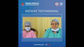
On 27th June 2008, I observed an unusual tumor on my right breast. I suspected it to be a cancer tumor & immediately went to Doctor and got mam.....

I was a 5 year old in 1997 when my cancer got detected. I started becoming very weak and often had blackouts and used to feel dizzy and fall down......
As told by her relative, when she was three years old she used to have pain in her stomach. It used to bloat, she used to have a high fever and not.....
In 1997 when I was teaching at Sophia school I came to know of a lump in my right breast. I went to SMS hospital and got it operated upon and had t.....
A person is known by his voice and was very worried when my voice got a little hoarse. I thought it may be because of a little change in the weathe.....
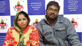
Babulaal Saini resident of Sanganer, Jaipur works as a laborer to fulfill the daily needs of his family. In 2005 after a painful period of 10 month.....
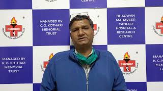
Mukesh Jain, a resident from Ajmer works in a marble factory. In the year 2016 he had felt a severe pain in the right side of his mouth.....
It was an evening of 2007, when I was playing with my son (Mohammed Azharuddin) and suddenly felt a lump on his neck. That time I didn’t took.....
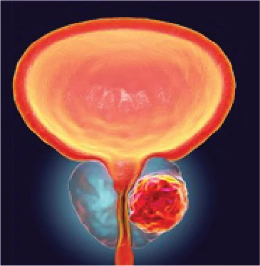
Radiology plays a critical role in Cancer diagnosis by utilizing advanced imaging techniques such as MRI, CT Scans, PET-CT Scans, and ultrasound to visualize and characterize tumors, aiding in their detection, localization, and staging.
Various imaging studies, including MRI, CT Scans, PET-CT Scans, ultrasound, and digital mammography, may be used depending on the type of cancer and the information needed for accurate diagnosis and treatment planning.
Interventional Oncology involves Minimally Invasive procedures performed under imaging guidance to diagnose, stage, and treat Cancer. These procedures include Tumor Ablation, Embolization, Biopsy, and other targeted therapies.
Minimally Invasive procedures offer advantages such as smaller incisions, reduced risk of complications, shorter recovery times, less postoperative pain, and improved cosmetic outcomes compared to traditional open surgeries.
Interventional Oncology procedures can be used to treat various types of Tumors, including Liver Tumors, Kidney Tumors, Lung Tumors, Bone Tumors, and soft tissue tumors.
Tumor Ablation involves the destruction of cancerous tissue using techniques such as Radiofrequency Ablation, Microwave Ablation, or Cryoablation. These procedures are performed under imaging guidance to precisely target and destroy the tumor while sparing surrounding healthy tissue.
TACE is a procedure used to treat liver tumors by delivering Chemotherapy drugs directly to the tumor site through the hepatic artery while simultaneously blocking the blood supply to the tumor, resulting in localized treatment and reduced systemic side effects.
Interventional Oncology procedures are generally considered safe when performed by experienced specialists in a controlled clinical setting. Risks and complications are minimized through careful patient selection and meticulous technique.
Recovery after Interventional Oncology procedures is typically quicker and less painful compared to traditional surgeries. Patients may experience mild discomfort, bruising, or fatigue, but most can resume normal activities within a few days to a week.
Results from Tumor Ablation or embolization procedures may vary depending on the type and size of the tumor, as well as the patient's overall health. In some cases, tumor shrinkage or resolution may be observed within weeks to months following the procedure.
Long-term effects or complications following Interventional Oncology procedures are rare but may include recurrence of the Tumor, damage to surrounding organs or tissues, or complications related to anesthesia or infection. Close monitoring and follow-up care are essential to detect and address any potential issues.
Yes, Interventional Oncology procedures can be combined with other Cancer treatments such as Surgery, Chemotherapy, Radiation Therapy, or Targeted Therapy, depending on the individual patient's diagnosis and treatment plan.
Preparation for an Interventional Oncology procedure may involve fasting before the procedure, discontinuation of certain medications, and following specific instructions provided by your healthcare team. It's essential to discuss any questions or concerns with your healthcare provider beforehand.
Most Interventional Oncology procedures are performed under local Anesthesia and sedation to minimize discomfort. Patients may feel some pressure or mild discomfort at the procedure site, but pain is generally well managed with medication and anesthesia.
During recovery, patients may experience mild discomfort, bruising, or fatigue, which can typically be managed with over-the-counter pain medications and rest. Your healthcare team will provide specific post-procedure instructions and follow-up care to ensure a smooth recovery process.
Request a callback from our healthcare specialist
 Careers
 Book an Appointment
 Patient Portal